SilverNanoparticlesOnEcoliBacteria.jpg
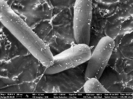
Photo by ZEISS Microscopy, Flickr / CC BY-NC-ND 2.0
E. coli, bacteria, cryo FIB-SEM, dotted with silver nanoparticles. Inlens image. Sample courtesy of R. Niessner, N. Ivleva, M. Seidel, A. Kunze, Institute of Hydrochemistry, Chair for Analytical Chemistry, TU Muenchen, Germany.
Introduction
Chronic Lyme disease patients are often afflicted with more than just borreliosis. The symptomatology and diagnoses of such patients is complicated, often involving multiple infections affecting every organ system in many cases. Such patients often suffer from chronic viral infections, multiple bacterial, parasitic, and protozoal infections, as well as chronic fungal and mold infections. Immune dysregulation, including reduced white blood cells and decreased natural killer cell activity, along with elevated inflammatory cytokines, biofilms, neurotoxins, and biotoxins often complicate the disease process further, providing a toxic milieu for disease progression.
Central to the current treatment approach is prescription antibiotic therapy, yet antibiotic therapy often causes bacterial resistance and may at best only suppress some of the bacterial infections while promoting the growth of biofilm formation, candida overgrowth, and disturbances in the gut microbiome, again contributing to the progression of the disease process. New resistant strains of bacteria, resulting from recurrent use of antibiotics, make antibiotic therapy less effective than previously thought.2 Such treatment strategies do not address all of the disturbances produced by the multiple infections; often multiple antibiotics are required to address the complexity of the infectious syndrome.
Hydrosol silver, specifically silver nanoparticles, provides a novel and effective approach to the treatment of chronic Lyme disease and its multiple infections without the complications of antibiotic therapy.
Chronic Lyme Disease, or Multiple Systemic Chronic Infection Syndrome (MSCIS)
Chronic Lyme disease, also known as multiple systemic chronic infection syndrome (MSCIS), involves multiple infections such as viruses, parasites, bacterial, spirochetes fungi, and mold. A combination of these infections and their toxins play a large role in furthering immune deficiency, increasing inflammation, damaging mitochondria, stimulating mTor and other pro-inflammatory protein enzymes, as well as causing endocrine, neurological, gastrointestinal, and psychoemotional disturbances.
Combination antibiotic therapy, to address the multitude of infection types, often leads to further inflammation, organ damage, extensive biofilm formation, neurotoxins, and biotoxins as well as promoting an acidic milieu perfect for the growth of mold, fungi, and parasitic infections. Silver nanoparticle therapy, on the other hand, provides a broad-spectrum antimicrobial therapy coupled with immune- and inflammation-modulating benefits as well as biofilm disruption capabilities, making it a more effective treatment for MSCIS.
History of Silver
Greeks, Romans, Phoenicians, and Egyptians used silver to preserve food and water, a household practice that continued until World War II. Persian kings, including Cyrus, consumed only drinking water carried in silver containers due to their ability to preserve its freshness. Hippocrates and Avicenna, the “father of modern medicine,” applied silver to wounds for healing and silver fillings for blood purification.10 For over six millennia, professionals and laypeople recognized the strong antimicrobial benefits of silver to eliminate a multitude of bacterial, parasitic, viral, and fungal infections. Topical dressings using silver nitrate to disinfect and heal open wounds, burns, and chronic ulcers continued to be utilized for centuries until the advent of antibiotics. Silver sulfadiazine was originally used for burn and wound victims during wars for its antibacterial and wound healing properties.1 Robert O. Becker, MD, in the 1970s, discovered the bone-growing properties and bactericidal effects of silver ions. Silver exhibits strong antimicrobial activity against a wide range of microorganisms.3
Silver Nanoparticles vs. Colloidal Silver
With the advent of nanotechnology, scientists developed silver nanoparticles, recognizing its potential in medicine and technology. Nano means “dwarf” in Greek and refers to the less than 100 nm atom size of the silver nanoparticles. Silver nanoparticles have a large surface area to volume ratio, making them more potent than colloidal or other forms of silver. Because of this large ratio, lower concentrations of silver nanoparticles exhibit potent antimicrobial activity against drug-resistant infections than higher concentrations of colloidal silver. Silver nanoparticles are bactericidal and bacteriostatic against multiple bacteria.6 Historical and scientific documentation verifies silver to be antiseptic against 650 different organisms including parasitic, viral, fungi, and mold infections.
During World War I, the primary form of silver was colloidal silver, which was used for wound healing and as antiseptic. The utilization of intravenous colloidal silver to treat major infections proved erroneous, as high doses of colloidal silver intravenously caused convulsions and deaths. High doses of oral colloidal silver caused gastrointestinal symptoms, resulting in the loss of routine use of silver as a mainstream antimicrobial therapy after the 1940s.
The introduction of nanotechnology and the development of silver nanoparticles welcomed silver back into medicine during the last few decades, as silver nanoparticles exhibited a significant reduction in toxicity compared with that of colloidal silver. Thus, silver nanoparticles display a high level of safety because of their large surface area to volume ratio. The introduction of silver nanoparticles eliminated the concerns with colloidal silver, including argyria (“blue man syndrome”), convulsions, and gastrointestinal side effects. Silver nanoparticles prove to be a stronger and more effective antimicrobial agent than colloidal silver without the toxicity or side effects. No cases of convulsions with silver nanoparticles have been reported, and because silver nanoparticles do not contain the salts and proteins of colloidal silver, the gastrointestinal side effects are eliminated. Chemically, silver nanoparticles are extremely stable and nonvolatile.
Due to the small size and large surface area, much smaller doses of silver nanoparticles are necessary for medical efficacy, further increasing safety. Silver nanoparticles produce the highest electrical and thermal conductivity of all metals; therefore, these nanoparticles have not only chemical activity against microbes but also thermal and electric activity.
Action of Silver Nanoparticles
Silver nanoparticles penetrate bacterial cell walls, causing structural changes to them, increasing membrane permeability, which leads to bacterial cell death. Silver nanoparticles prevent cell division and DNA replication and possess inhibitory and bactericidal effects against gram negative and gram positive bacteria, including Streptococcus pneumoniae, Staphylococcus aureus, and Enterococcus faecalis.
Through modulating the phosphotyrosine profile and interfering with transduction signaling of bacteria, silver nanoparticles inhibit bacterial growth.5 Inhibition of biofilm formation prevents drug-resistant bacteria, as silver nanoparticles disrupt the cytoskeleton of bacteria.7 Biofilm formation of Pseudomonas aeruginosa and Escherichia coli, for example, and other gram negative bacteria are inhibited and disrupted by exposure to silver nanoparticles.9 Brushing teeth with silver nanoparticles reduced the total and number of bacterial genome of supra- and subgingival more effectively than chlorhexidine.20
As an antifungal, silver nanoparticles cause die-off of multiple strains of Candida, including C. albicans, C. glabrata, C. parapsilosis, and C. krusei, as well as Trichophyton,which causes jock itch, ringworm, and toenail fungus. By disrupting cell membrane structure and inhibiting the normal budding process of fungi, silver nanoparticles destroy fungi habitation, healing fungal-related conditions. Through cell-wall disruption of the fungi, including Candida albicans, silver nanoparticles strongly inhibit their biofilm formations.8
Silver nanoparticles also have strong antiviral capabilities, lowering counts of viruses including HIV, herpes, RSV, hepatitis B, and multiple others. The mechanism of action of silver nanoparticles on viruses comprises glycoprotein binding, thereby inhibiting viral replication at early onset of the infection.22
As a potent tool against parasitic infections, silver nanoparticles have proved effective against Entamoeba histolytica, Cryptosporidium parvum, and Plasmodium falciparum, the intracellular parasite causing malaria.11,12 Babesia microti, another intracellular parasite, behaves similarly to Plasmodium falciparum, is resistant to most antimicrobial therapy, and is regarded as a coinfection of chronic Lyme disease. Since silver nanoparticles destroy Plasmodium falciparum, which resembles Babesia microti, the possibility that silver nanoparticles may be also efficacious in the treatment of Babesia microti is plausible despite the lack of current research.
Biofilm and Silver Nanoparticles
Biofilm is the “cocoon” or gelatin that microbes create around themselves to prevent immunological attacks, providing self-protection. Microbes form complex networks of biofilm, such as dental plaques, which are difficult to eradicate, thus allowing the microbes to hide from both antibiotic therapy and the immune system, making diagnosis and treatment of intracellular infections such as babesiosis and borreliosis difficult.
Unlike antibiotic therapy, which encourages an increase in biofilm formation, silver nanoparticles interfere with biofilm development, reducing the virulence of infections.18 Silver nanoparticles, furthermore, enhance drug or herbal transport to the infections, acting as delivery a system to the biofilm. For example, when silver nanoparticles merge with curcumin, an extract of turmeric, biofilm destruction is significantly potentiated.11
Many of the virulent bacteria, such as Borrelia burgdorferi, produce endotoxins and biotoxins that further damage joints, tissues, and organs, often causing pain and inflammation. Recent research by Lambadi et al. confirms not only the biofilm control but the removal of endotoxins by silver nanoparticles, illustrating further advantage of silver nanoparticles as a promising treatment for chronic Lyme disease and its associated infections.18
Other Effects of Silver Nanoparticles
Modulation of pro-inflammatory cytokines, such as IL-2, IL-6, TNF-α, by silver nanoparticles in cancer cells, specifically in leukemic cells and in infections, contributes further to potential benefits in patients with cancer and concurrent chronic Lyme disease.17 Further studies are needed to assess silver nanoparticles’ effect on pro- inflammation of monocytes, since they may also upregulate pro-inflammatory cytokines, as shown in one study.19 Silver nanoparticles used in mice temporarily disturbed the intraendothelial junction of cells and induced mitochondrial stress, which was corrected with combination therapy with sodium selenite, or selenium supplementation.14-16
As the microbiome controls approximately 70% of the immune system, the role of oral silver nanoparticles in disturbing the gut microbiome must be considered. Fortunately, silver nanoparticles have a dose-dependent effect on the Lactobacillus genus and the Firmicutes population in rats, implying that careful oral dosing of silver nanoparticles becomes strategically important in protecting the microbiome.21 Alternatively, the prudent use of a nutrition and dietary supplementation optimizing pre- and probiotics concomitantly with silver nanoparticle oral dosing is worthy in maintaining proper microbiome balance and protection.
Clinical Cases Observed
A 55-year-old female with chronic pain syndrome, fibromyalgia, neuropathy, and a diagnosis of chronic Lyme disease with multiple infections was treated with two rounds of intravenous and oral hydrosol silver for 4 weeks. Symptoms of chronic pain, neuropathy, and inflammation significantly reduced, and the patient was able to return to functional status.
A 46-year-old female with a diagnosis of interstitial cystitis caused by chronic Lyme disease was given two rounds of a moderate dose of intravenous and oral hydrosol silver. Her chronic interstitial cystitis resolved within a few weeks of initiating treatment.
A 52-year-old female with chronic Lyme disease with presenting symptoms of brain fog and chronic sinusitis had her chronic symptoms resolve upon nebulizing hydrosol silver.
Conclusion
Silver nanoparticles provide strong antimicrobial therapy against hundreds of microbes, including intracellular bacteria, parasites, fungi, and viruses. By disrupting biofilm formation, reducing endotoxins, and destroying microbial replication and virulence, silver nanoparticles play a powerful role in the complex treatment of chronic Lyme disease and associated infections, often reducing clinical symptoms and improving physical function. Though silver nanoparticles have clinically proved to be very safe, further research is needed to understand the immunological modulation of silver nanoparticle on humans fighting chronic infections. In the meantime, to minimize any possible adverse reactions from oral or intravenous use of hydrosol or silver nanoparticles, selenium or sodium selenite along with probiotics should be considered.
Notes
- Feng QL, Wu J, Chen GQ, Cui FZ, Kim TN, Kim JO. A mechanistic study of the antibacterial effect of silver ions on Escherichia coli and Staphylococcus aureus. J Biomed Mater Res. 2000;52:662–668.
- Kyriacou SV, Brownlow WJ, Xu X‐H. Using nanoparticle optics assay for direct observation of the function of antimicrobial agents in single live bacterial cells. Biochemistry. 2004;43:140–147.
- Liau SY, Read DC, Pugh WJ, Furr JR, Russell AD. Interaction of silver nitrate with readily identifiable groups: relationship to the antibacterial action of silver ions. Lett Appl Microbiol. 1997;25:279–283.
- Kirstein J, Turgay K. A new tyrosine phosphorylation mechanism involved in signal transduction in Bacillus subtilis. Mol Microbiol Biotechnol. 2005;9:182.
- Morones JR et al. The bactericidal effect of silver nanoparticles. Nanotechnology. 26 August 2005;16(10).
- Sanyasi S et al. Polysaccharide‐capped silver Nanoparticles inhibit biofilm formation and eliminate multi‐drug‐resistant bacteria by disrupting bacterial cytoskeleton with reduced cytotoxicity towards mammalian cells. Sci Rep. 2016 Apr 29;6:24929. doi:10.1038/srep24929.
- Lara HH et al. Effect of silver nanoparticles on Candida albicans biofilms: an ultrastructural study. J Nanobiotechnol. 2015 Dec 15;13:91. doi:10.1186/ s12951‐015‐0147‐8.
- Radzig MA et al. Antibacterial effects of silver nanoparticles on gram‐negative bacteria: Influence on the growth and biofilms formation, mechanisms of action. Colloids Surf B Biointerfaces. 1 February 2013;102:300–306.
- Alexander WJ. History of the medical use of silver. Surg Infect. 2009;10(3).
- Loo CY et al. Combination of silver nanoparticles and curcumin nanoparticles for enhanced anti‐biofilm activities. J Agric Food Chem. 2016 Mar 30;64(12):2513–2522. doi:10.1021/acs.jafc.5b04559. Epub 2015 Dec 3.
- Saad HA, Soliman MI, Azzam AM, Mostafa B. Antiparasitic activity of silver and copper oxide nanoparticles against Entamoeba histolytica and Cryptosporidium parvum cysts. J Egypt Soc Parasitol. 2015 Dec;45(3):593–602.
- Jaganathan A et al. Earthworm‐mediated synthesis of silver nanoparticles: a potent tool against hepatocellular carcinoma, Plasmodium falciparum parasites and malaria mosquitoes. Parasitol Int. 2016 Jun;65(3):276–284. doi:10.1016/j.parint.2016.02.003. Epub 2016 Feb 9.
- Guo H et al. Intravenous administration of silver nanoparticles causes organ toxicity through intracellular ROS‐related loss of inter‐endothelial junction. Part Fibre Toxicol. 2016 Apr 29;13:21. doi:10.1186/s12989‐016‐0133‐9.
- Teodoro JS et al. Low‐dose, subchronic exposure to silver nanoparticles causes mitochondrial alterations in Sprague‐Dawley rats. Nanomedicine (Lond). 2016 May 12.
- Ma W, Jing L. Silver nanoparticle exposure induced mitochondrial stress, caspase‐3 activation and cell death: amelioration by sodium selenite. Int J Biol Sci. 2015 Jun 1;11(8):860–867. doi:10.7150/ijbs.12059. eCollection 2015.
- Parnsamut C, Brimson S. Effects of silver nanoparticles and gold nanoparticles on IL‐2, IL‐6, and TNF‐α production via MAPK pathway in leukemic cell lines. Genet Mol Res. 2015 Apr 17;14(2):3650–68. doi:10.4238/2015.April.17.15.
- Lambadi et al, Facile biofunctionalization of silver nanoparticles for enhanced antibacterial properties, endotoxin removal, and biofilm control. Int J Nanomedicine. 2015 Mar 18;10:2155–2171. doi:10.2147/IJN.S72923. eCollection 2015.
- Murphy A et al. Silver nanoparticles induce pro‐inflammatory gene expression and inflammasome activation in human monocytes. J Appl Toxicol. 2016 Mar 10. doi:10.1002/jat.3315.
- Do Nascimento C et al. Microbial diversity of the supra‐ and subgingival biofilm of healthy individuals after brushing with chlorhexidine‐ or silver‐coated toothbrush bristles. Can J Microbiol. 2015 Feb;61(2):112–123. doi:10.1139/cjm‐2014‐0565. Epub 2014 Nov 5.
- Williams K, Milner J, Boudreau MD, Gokulan K, Cerniglia CE, Khare S. Effects of subchronic exposure of silver nanoparticles on intestinal microbiota and gut‐associated immune responses in the ileum of Sprague‐Dawley rats. Nanotoxicology. 2015 May;9(3):279–289. doi:10.3109/17435390.2014.921346. Epub 2014 May 30.
- Hu RL, Li SR, Kong FJ, Hou RJ, Guan XL, Guo F. Inhibition effect of silver nanoparticles on herpes simplex virus 2. Genet Mol Res. 2014 Mar 19;13(3):7022–7028. doi:10.4238/2014.March.19.2.
Silver Nanoparticles as a Novel Approach to the Treatment of Chronic Lyme Disease and Associated Diseases was originally published by Townsend Letter, June 10, 2022. Used with permission.